Advanced Imaging Center (BSL2/BSL3)
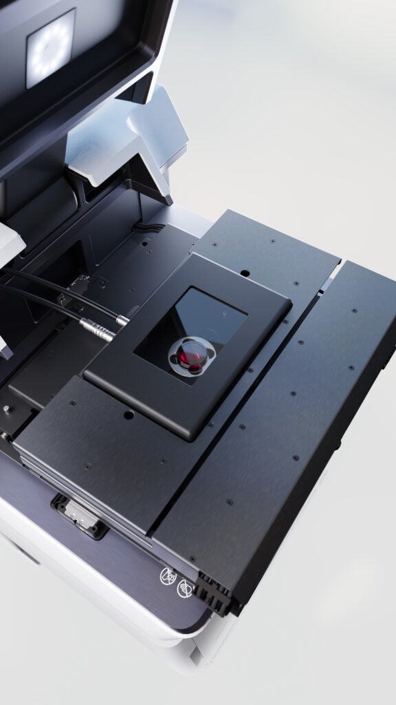
The IID-HSPH BSL-3 Imaging Core, established in 2024 with funding from the Massachusetts Life Sciences Center, provides advanced fluorescence microscopy capabilities for imaging biological samples across BSL-2 to BSL-3 containment environments. The facility serves researchers at the Harvard T.H. Chan School of Public Health and the broader Massachusetts scientific community, offering access to state-of-the-art live-cell Spinning Disk Confocal and Lattice Light Sheet microscopes.
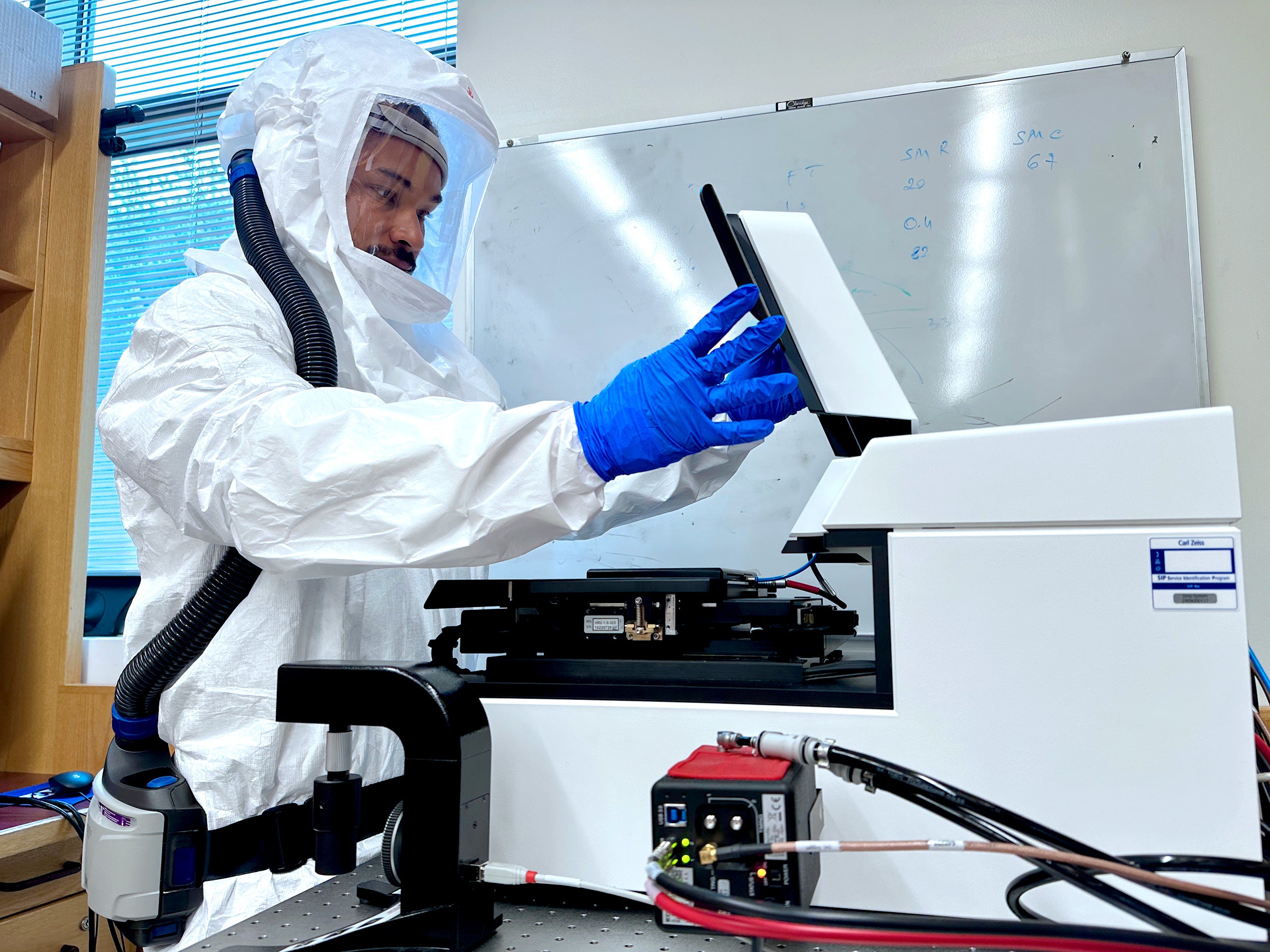
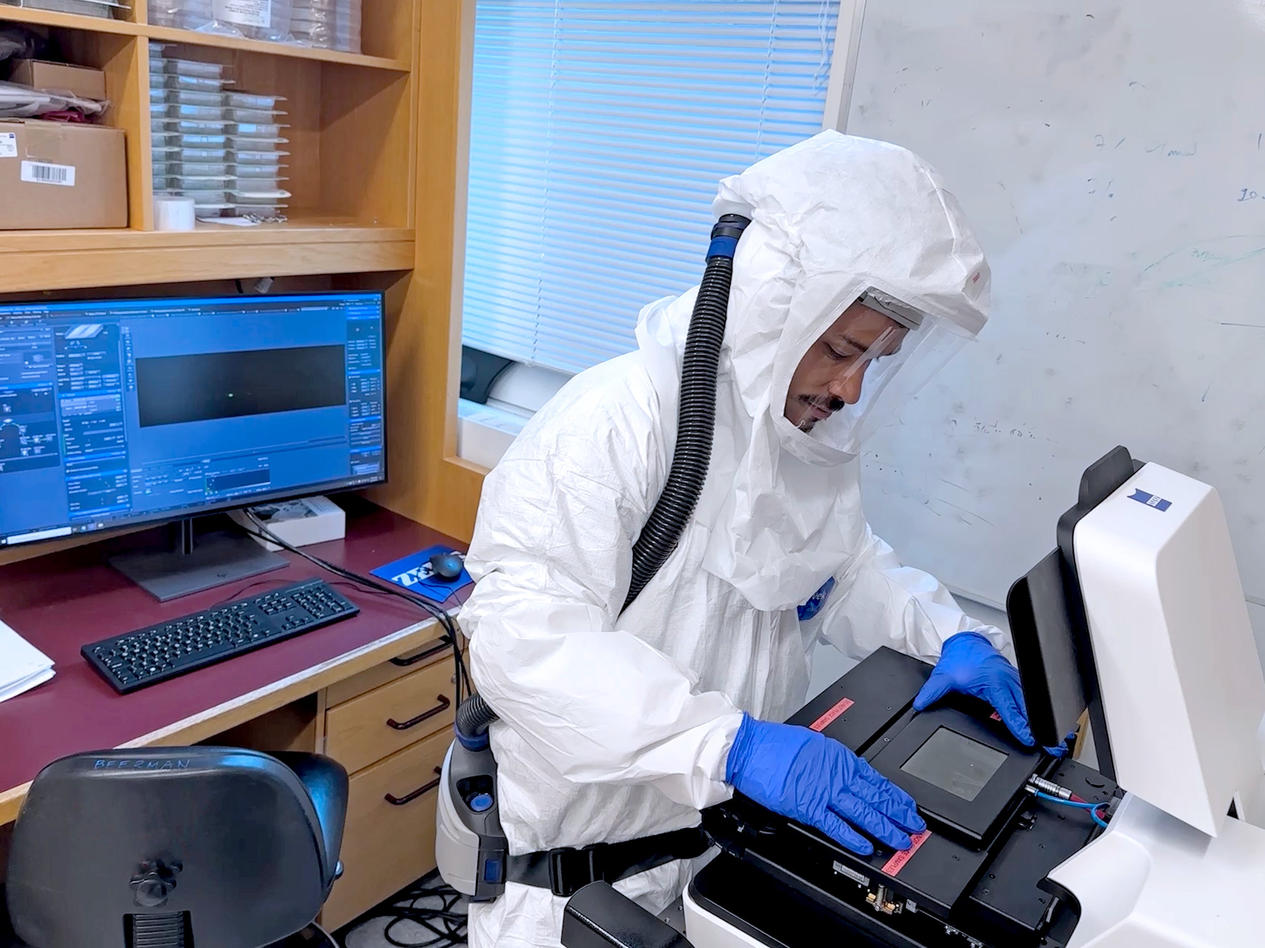
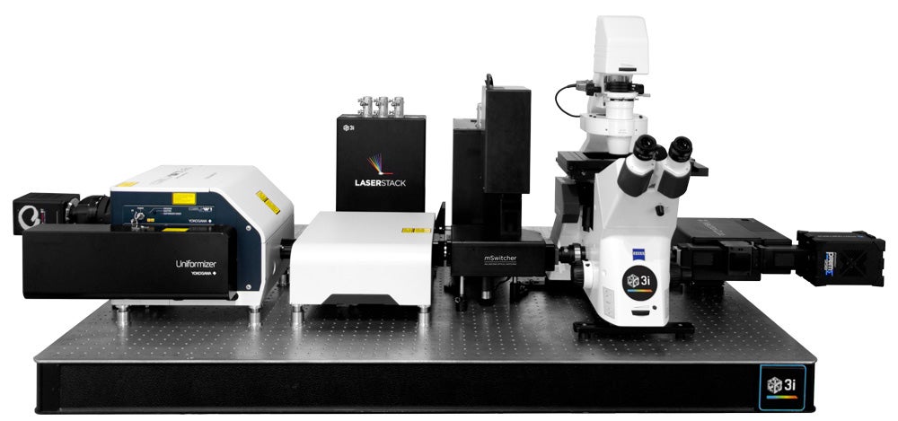
Spinning Disk Confocal
Microscope
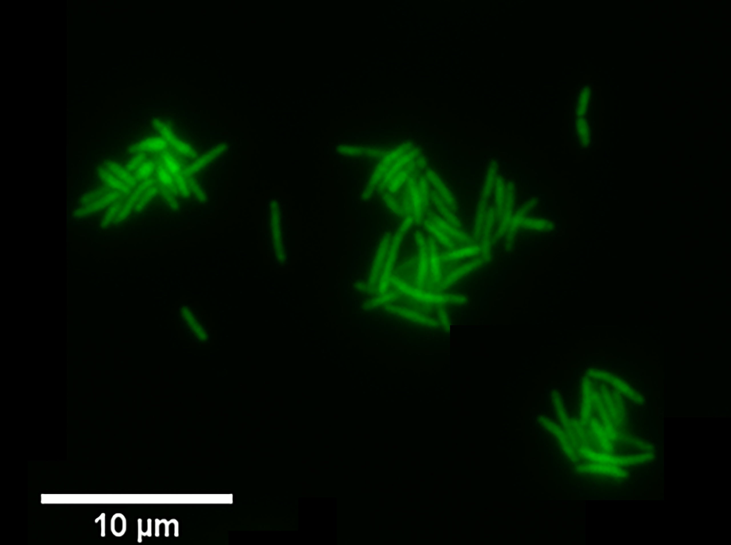
This system enables high-speed, high-resolution imaging of both fixed and live cells and tissues. With lateral (XY) and axial (Z) resolutions of approximately 300 nm and 700-1000 nm, respectively, it is well-suited for multichannel fluorescence imaging in a BSL-3 context. Key features include:
- 40x Oil, NA 1.3
- 63x Oil, NA 1.4
- 10x Air, NA 0.45
- 20x Air, NA 0.8
Excitation Lasers: 488 nm, 561 nm, 638 nm
Detection: Dual sCMOS cameras for simultaneous two-channel acquisition
This configuration supports rapid volumetric imaging with minimal phototoxicity and is optimized for dynamic cellular processes.
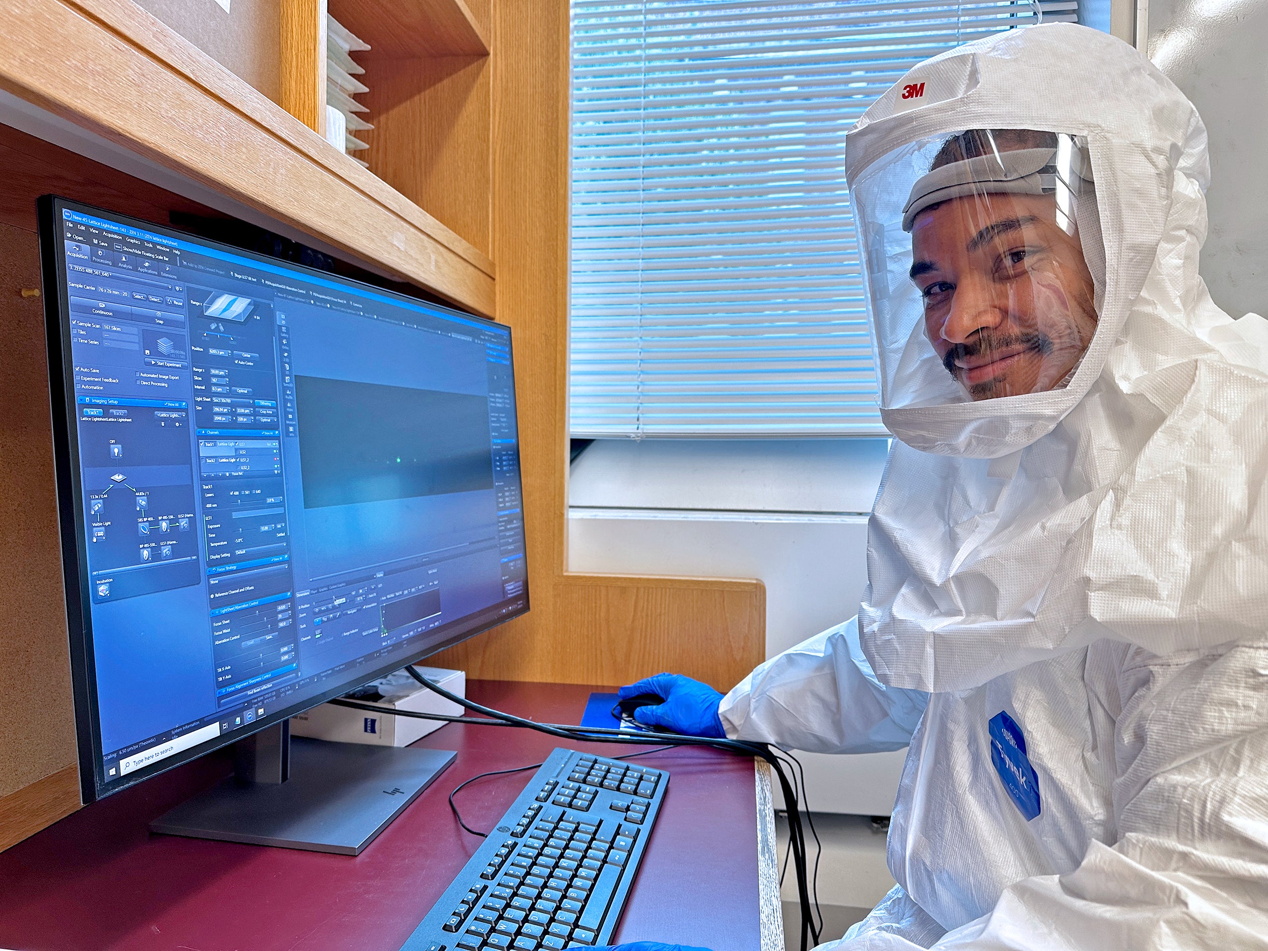
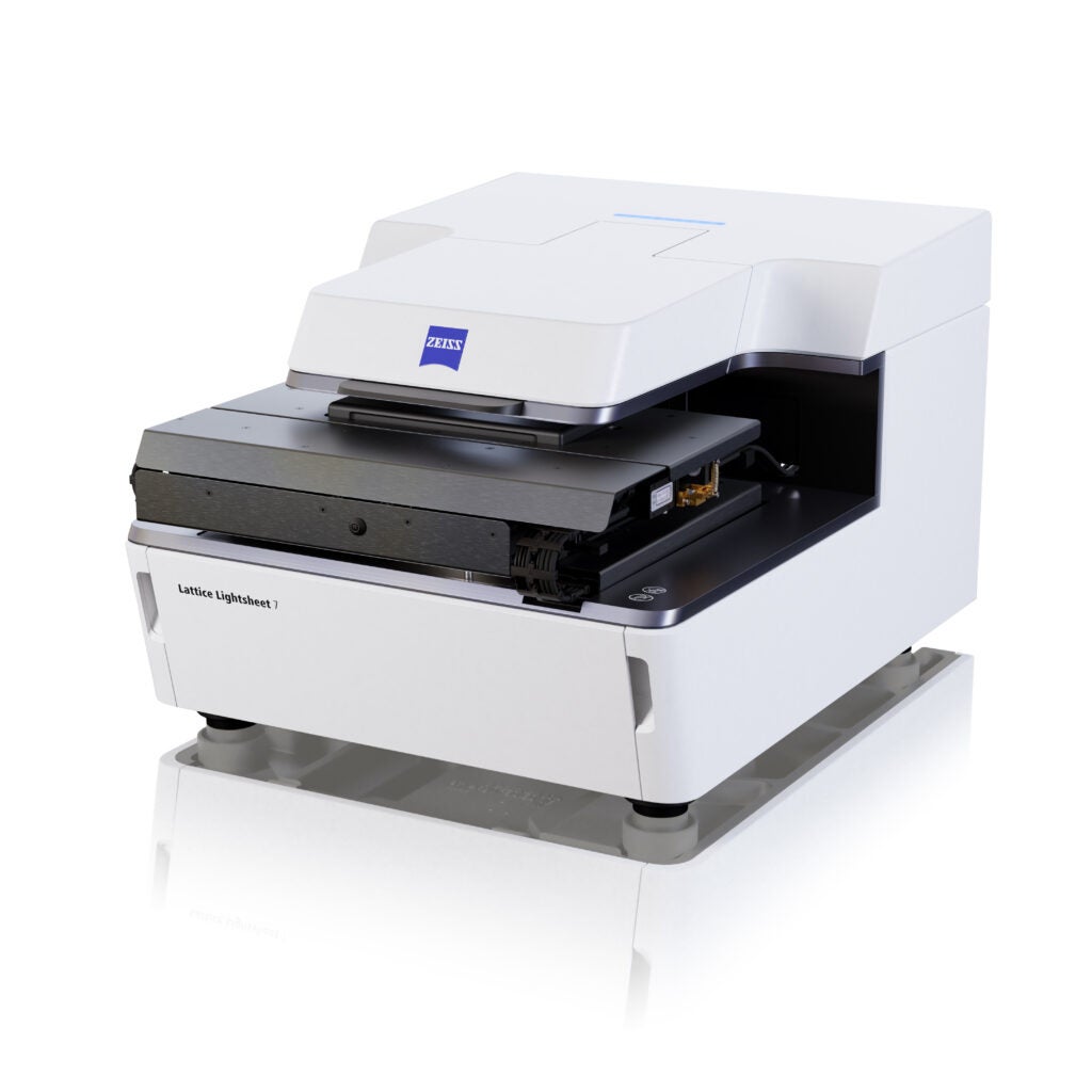
Lightsheet Miscroscope
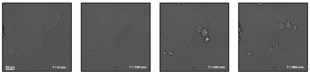
The Zeiss Lattice Lightsheet 7 enables high-resolution, low-phototoxicity imaging of live cells and tissues. It combines lattice light sheet illumination with single-objective detection for gentle (at least 100 times better than the spinning disk confocal microscope if imaging 100 planes of a cell), volumetric imaging over time. The system achieves lateral and axial resolutions of approximately 300 nm and 600 nm, respectively.
Key Features:
Objective: 44.83x, NA 1.0 (single-objective configuration)
Excitation Lasers: 488 nm, 561 nm, 640 nm
Detection: Dual sCMOS cameras for simultaneous multichannel acquisition
This system is ideal for time-resolved 3D imaging of dynamic cellular events with minimal photobleaching and photodamage.
Access and Training
To use the microscopy systems, please first contact Dr. Ricardo F. Bango Da Cunha Correia to discuss your scientific goals and experimental design. Following this consultation, tailored training will be provided based on your experimental needs. Access to the microscopes can be scheduled once training is complete.
Data Management
For external users: Please bring your own external hard drive for data collection. A dedicated workstation is available on-site for data visualization and analysis.
Location and Contacts
The BSL3 core Facility is located in the Francois-Xavier Bagnoud Building (FXB).
For more information or to schedule an appointment, please contact the BSL-3 Imaging Director, Ricardo Bango Da Cunha Correia.
Ricardo Bango da Cunha Correia
Research Associate
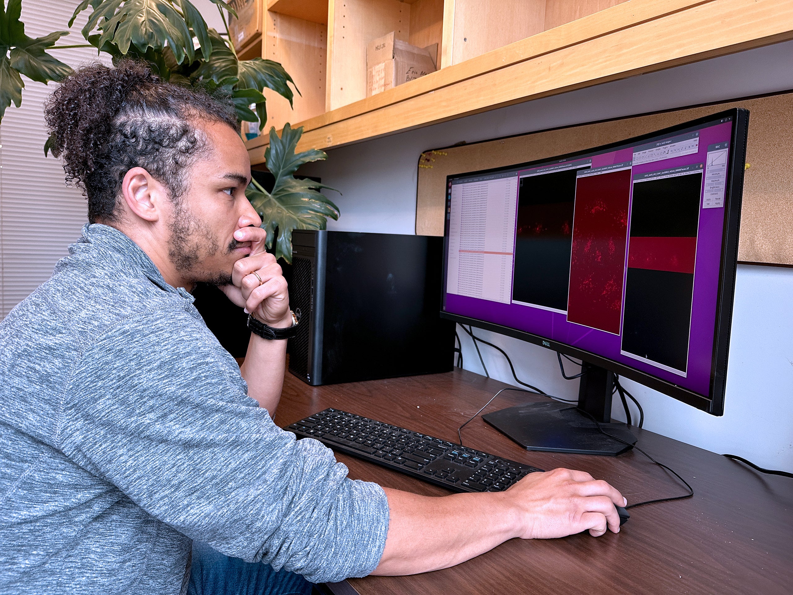
Ricardo has joined IID to manage the advanced microscopy facility that is currently being implemented in the department. He is a Physicist with a PhD in Life Sciences which he earned in Dundee, Scotland. He has more than 10 years of experience in designing, building and operating microscopes with emphasis on Lightsheet and Lattice Lightsheet microscopy.
During the last 6 years Ricardo has worked in Tom Kirchhausen’s Lab at BCH/HMS, where he built a MOSAIC scope and conducted live imaging experiments with cells, tissues, drug discovery and viral infection. In some of the experiments microfluidic devices were designed and implemented to monitor and time, as an example, cell-cell interaction.
The BSL3 core Facility is located in the Francois-Xavier Bagnoud Building (FXB).
If you need more information or you’d like to schedule an appointment please contact the BSL3 Imaging Director, Ricardo Bango Da Cunha Correia.

Noman Siddiqi
Director of Research Operations
Noman is the Director of Research Operations in the department.